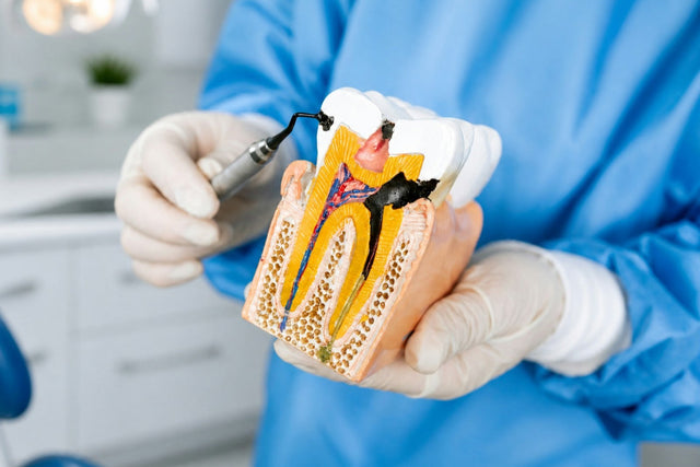
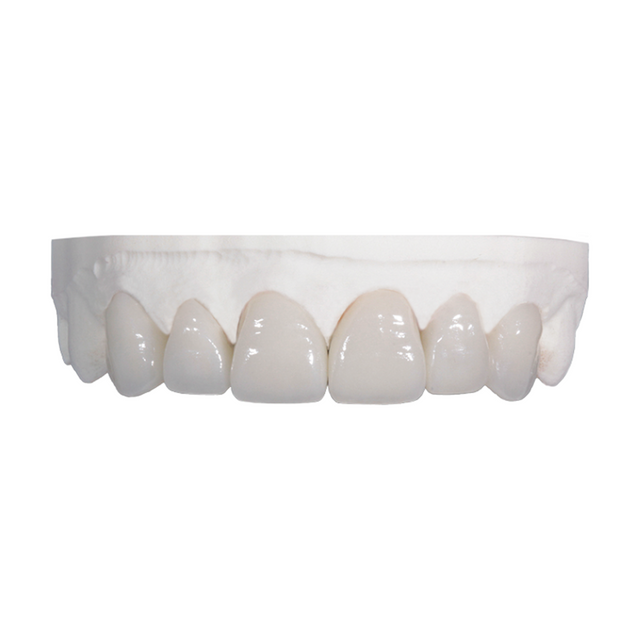
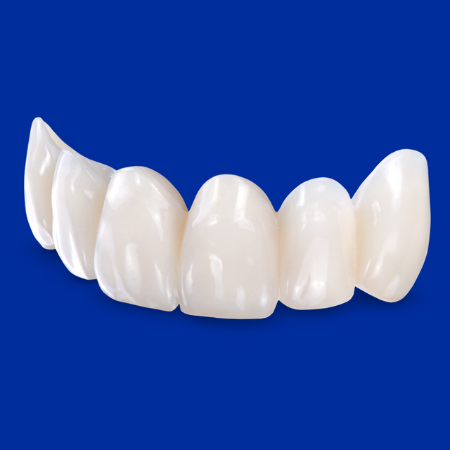
A molar comes in with a failing MOD amalgam. With the filling out, the buccal cusps look sound, the lingual cusp is thin, and the patient asks whether the tooth really needs a crown. A crown is the default answer; a bonded onlay would keep far more of the tooth.
Below: what each preparation removes, how long each option lasts, when a cusp needs coverage, and how to prep, bond and prescribe IPS e.max inlays and onlays.
Key Takeaways
- In a 2025 digital molar study, a bonded IPS e.max onlay removed 16.9% of the crown, a partial crown 27.3% and a full crown 55.3%.
- Ceramic inlays and onlays survived 92% to 95% at 5 years and 91% at 10 years in a meta-analysis of 14 studies.
- Crowns set a high bar: 98.5% five-year survival for monolithic lithium disilicate and 97.1% for metal-ceramic in a 2026 meta-analysis.
- Cover cusps thinner than about 2 mm: in a lab study, 1.0 mm cusp walls cracked more than 2.0 mm walls under ceramic inlays.
- In a 7-year study of painful cracked teeth, restorations without cusp coverage failed at 6% a year; none with cusp coverage failed.
Quick Answer
Choose an inlay when the cusps are sound and at least about 2 mm thick; an onlay when a cusp is thin, cracked or missing but enough enamel remains to bond; and a full crown when little coronal structure is left, the margins cannot be kept dry for bonding, or the patient has heavy parafunction. In the studies available, bonded ceramic onlays survive about as well as crowns and remove far less tooth.
How Much Tooth Does Each Preparation Remove?
Weighing resin typodont teeth before and after preparation, Edelhoff and Sorensen found that adhesive and inlay preparations removed about 5.5% to 27.2% of tooth structure, the MOD inlay at the top of that range, while complete crowns removed 67.5% to 75.6%.
A 2025 study modeled preparations on a digital maxillary first molar at manufacturers' minimum thicknesses. With IPS e.max CAD bonded at 1.0 mm, an occlusal onlay removed 16.9% of the crown volume, a partial crown 27.3% and a full crown 55.3%. An e.max crown at the 1.5 mm needed for self-adhesive or conventional cement took 65.1%, and a monolithic zirconia crown at 0.5 mm still took 43.0%. Finish-line distance from the CEJ mattered most, so keep margins as far from it as the lesion allows.
Share of a molar crown removed for a bonded e.max occlusal onlay, against 27.3% for a partial crown and 55.3% for a full crown, all at 1.0 mm (Seidel et al., 2025).

Do Inlays and Onlays Last as Long as Crowns?
Ceramic inlays and onlays
A 2016 meta-analysis of 14 studies with more than 5 years of follow-up put survival of glass-ceramic and feldspathic inlays, onlays and overlays at 92% to 95% at 5 years (5,811 restorations) and 91% at 10 years (2,154). Fracture or chipping was the most common failure (4%), ahead of endodontic complications (3%). In one private practice, 556 pressed IPS e.max partial-coverage restorations reached 95.6% cumulative survival at 10 years, and all 6 failures were in molars. In the same group's 16.9-year cohort, 6 of 610 partial-coverage and 16 of 1,782 complete-coverage posterior restorations failed.
Gold and composite
Cast gold has the longest records. In a university study, IPS Empress inlays and onlays matched cast gold partial crowns at 23.5 years (91.7% vs. 91.8%); the ceramics' most common complication was fracture (6.7%), the gold's endodontic complications (8.8%). Composite trails: indirect composite had an 18% higher failure rate than intracoronal gold over 5 to 7 years, and composite onlays pooled 90% survival against 98% for lithium disilicate.
Full crowns and head-to-head data
Single crowns have strong five-year data (see the table), but direct comparisons are few. A 2022 meta-analysis found no significant survival difference between onlays or partial crowns and full crowns at 1 and 3 years. In a review of MOD-prepared teeth, all three eligible studies described crown failures as more catastrophic, while teeth with onlays could be salvaged.
| Restoration | Evidence | Survival | Follow-up |
|---|---|---|---|
| Glass-ceramic and feldspathic inlays, onlays, overlays | Meta-analysis, 14 studies | 92% to 95%; 91% | 5 years; 10 years |
| Pressed e.max inlays (246) | Private-practice cohort | 93.9% | 9.9 years |
| Pressed e.max onlays (305) | Same cohort | 98.3% | 9.8 years |
| Lithium disilicate onlays; composite onlays | Meta-analysis, 27 studies | 98%; 90% | Studies of 2 to 15 years |
| Cast gold inlays and onlays (303) | Cohort | 96.1%; 87.0%; 73.5% | 10; 20; 30 years |
| IPS Empress inlays and onlays vs. cast gold partial crowns | University cohort | 91.7% vs. 91.8% | 23.5 years |
| Single crowns: monolithic lithium disilicate; metal-ceramic; monolithic zirconia | Meta-analysis, 64 studies | 98.5%; 97.1%; 96.8% | 5 years |

When Does a Tooth Need an Inlay, an Onlay or a Crown?
Lesion size and missing cusps
Bustamante-Hernández et al. list a wide isthmus (over a third of the occlusal width), extensive proximal loss and lost cusps as reasons to go indirect. A lost cusp makes it an onlay.
The 2 mm cusp rule
In extracted molars with bonded ceramic inlays, non-functional cusp walls left 1.0 mm thick formed significantly more enamel cracks after thermal and mechanical loading than 2.0 mm walls; the authors recommend at least 2.0 mm. After caries removal, if a cusp wall is thinner than about 2 mm, reduce it and cover it.
Cracked teeth
In a 7-year study of painful cracked teeth restored with direct composite, restorations without cuspal coverage failed at a mean 6% per year and none with coverage failed. Bonded indirect composite onlays on 43 cracked teeth had 93.02% survival at 6 years. In the National Dental PBRN's Cracked Tooth Registry, dentists recommended a complete crown for 61% of the teeth they planned to restore and a partial crown for 3%. Full or partial coverage, especially full, succeeded more often than intracoronal restorations, and the authors call a full crown the most successful treatment.
For cracked teeth in more detail, see Cracked Tooth Restoration: Onlay, Crown or Watch?
Root-canal-treated teeth
In a University of Iowa cohort, root-filled teeth not crowned after obturation were lost at 6.0 times the rate of crowned teeth, and an IAAD consensus review weakly favored indirect restorations, mostly crowns, over direct ones. Nonvital teeth fail more under partial coverage: a review of ceramic onlays linked nonvital teeth and parafunction to more failures. A 2024 review found comparable survival for bonded partial restorations and crowns on root-filled posterior teeth but called the evidence limited. Cover every cusp; if the walls are thin, crown it.
Esthetics
Ivoclar lists three drivers of the final shade: the prepared tooth, the restoration (shade, translucency, thickness) and the cement, so a thin e.max onlay over a dark tooth needs a stump shade. Gold lasts but shows; authors of a 1,314-restoration gold study suggest it for patients more concerned with longevity than esthetics.
Which Restoration Fits Which Situation?
| Situation | Best option | Why |
|---|---|---|
| Moderate MOD, cusp walls about 2 mm or thicker | IPS e.max inlay | Least tooth removed; 2.0 mm walls cracked less than 1.0 mm walls |
| One cusp thinner than about 2 mm, cracked or missing | Onlay covering that cusp | Protects the weak cusp and keeps the rest |
| Wide isthmus, several weak cusps, vital tooth | Onlay covering all cusps | 16.9% to 27.3% removed, against 55.3% for a bonded full crown |
| Symptomatic cracked tooth | Cusp coverage: onlay or crown | No failures with coverage vs. 6% a year without; registry favors crowns |
| Root-canal-treated posterior tooth | All cusps covered; crown if walls are thin | Uncrowned root-filled teeth lost at 6.0 times the rate |
| Heavy bruxer, large defect | Monolithic full crown | Parafunction linked to more onlay failures; see restorations for bruxers |
| Margins cannot be kept dry | Crown on a retentive prep | E.max inlays and onlays must be bonded |
| Visible margins, esthetic demand | IPS e.max inlay or onlay | Tooth-colored margins and shade control |
How Should You Prepare for an E.max Inlay or Onlay?
Ivoclar's current IPS e.max CAD and Press instructions (2026) and its IPS e.max Clinical Guide give these rules:
- At least 1.0 mm preparation depth and 1.0 mm isthmus width in the fissure area.
- Onlays and partial crowns: at least 1.0 mm of space over the cusps. Partial crowns add a circular shoulder with rounded inner edges, or a chamfer of about 10° to 30°, at least 1.0 mm wide.
- Proximal boxes with slightly diverging walls (6°) and an angle of 100° to 120° between the cavity wall and the future proximal surface.
- Rounded internal edges and transitions, to prevent stress concentration in the ceramic.
We also advise no slice cuts or feather edges, and no margins on centric contacts. Keep ceramic margins as butt joints: in a 2023 lab study of CAD/CAM inlays, 90° margins gave IPS e.max CAD a significantly smaller marginal gap than 135° bevels. Crown reductions are in our Crown Prep Guide. Treat 1.0 mm as a floor: a systematic review associated longer onlay survival with at least 2 mm of occlusal ceramic, although in Malament's cohort thickness did not affect survival.
How Do You Bond E.max Inlays and Onlays?
Ivoclar makes adhesive cementation mandatory for e.max inlays, onlays and partial crowns; conventional and self-adhesive cements are for crowns at least 1.5 mm thick on a retentive preparation. So if you cannot keep the margins dry, choose a crown. A 2021 Cochrane review found low-certainty evidence, from direct composite restorations, that rubber dam improved six-month survival (odds ratio 2.29).
For the restoration, Ivoclar specifies IPS Ceramic Etching Gel followed by Monobond Plus, or one-step Monobond Etch & Prime, and says e.max CAD must never be blasted with alumina. Immediate dentin sealing is optional: meta-analyses of lab studies found higher microtensile bond strength with it, on low-quality evidence, but no difference in marginal gaps or microleakage. Etch and silane times are in How to Cement Zirconia and Bond E.max Crowns.
What Should You Send the Lab?
- Scan or impression: the whole preparation with every margin visible, full adjacent teeth, the opposing arch and a bite on both sides (How to Send a Scan to a Dental Lab).
- Design: inlay or onlay, which cusps to cover, and pressed or milled e.max if you have a preference (E.max Press vs. E.max CAD).
- Contacts and occlusion: your contact preference, and a note if an opposing cusp lands near a margin.
- Shade: tooth shade plus the shade of the prepared tooth, which Ivoclar reads at the largest, most discolored area (Shade Matching for the Lab).
- Codes: ceramic inlays are D2610 to D2630 and ceramic onlays D2642 to D2644, by number of surfaces (CDT Codes for Dental Lab Work).

Inlays and Onlays at Elegant Dental Laboratory
- We make IPS e.max inlays, onlays and occlusal onlays (table tops), cast gold inlays and onlays with an alloy certificate, and full-contour and layered crowns and veneers, every case in house in Brooklyn, with more than 35 technicians in a 7,000 sq. ft. facility.
- Digital scans are reviewed before production. Turnaround is from 5 business days on most restorative cases; rush is decided case by case.
- Up to a 7-year warranty on final restorations (policies and warranties).
- Submit a digital case or send a case: our own drivers pick up free every business day in the five NYC boroughs and 13 northern New Jersey counties, and UPS labels work anywhere. Call 877-335-5221.
Related Guides
- Zirconia vs. E.max: When to Use Each, With Prep and Cementation Tips
- Why Crowns Come Back High or With Open Contacts, and How to Prevent It
- 3-Unit Bridge vs. Implant Crown in 2026: What the Evidence Says
FAQ
Does an onlay last as long as a crown?
In head-to-head studies, yes, at least short term: no significant survival difference at 1 and 3 years in a 2022 meta-analysis. Failed onlays also leave a more salvageable tooth.
How thick must an e.max onlay be?
At least 1.0 mm in the fissure and over the cusps, per Ivoclar, and it must be bonded.
When should a cusp be covered?
When the remaining wall is thinner than about 2 mm, or the cusp is cracked or missing.
Can I seat an e.max inlay with RMGI or self-adhesive cement?
No. Ivoclar lists adhesive cementation only for e.max inlays, onlays and partial crowns.
Does a root-canal-treated molar always need a crown?
It needs cusp coverage. Crowns have the most data; an onlay covering all cusps can work when the walls are sound, though nonvital teeth fail more under partial coverage. On molars, an endocrown is another option; see our endocrown guide.
Sources (29)
- Edelhoff D, Sorensen JA. Tooth structure removal associated with various preparation designs for posterior teeth. Int J Periodontics Restorative Dent. 2002;22(3):241-249. PubMed 12186346
- Seidel K, et al. Quantitative analysis of substance removal during tooth preparation for full ceramic restorations using digitally generated preparation designs. Clin Oral Investig. 2025;29(6):298. doi:10.1007/s00784-025-06370-w
- Morimoto S, et al. Survival rate of resin and ceramic inlays, onlays, and overlays: a systematic review and meta-analysis. J Dent Res. 2016;95(9):985-994. doi:10.1177/0022034516652848
- Bustamante-Hernández N, et al. Clinical behavior of ceramic, hybrid and composite onlays: a systematic review and meta-analysis. Int J Environ Res Public Health. 2020;17(20):7582. doi:10.3390/ijerph17207582
- Malament KA, et al. 10.9-year survival of pressed acid etched monolithic e.max lithium disilicate glass-ceramic partial coverage restorations. J Prosthet Dent. 2021;126(4):523-532. doi:10.1016/j.prosdent.2020.07.015
- Malament KA, et al. Comparison of 16.9-year survival of pressed acid etched e.max lithium disilicate glass-ceramic complete and partial coverage restorations in posterior teeth. J Prosthet Dent. 2021;126(4):533-545. doi:10.1016/j.prosdent.2020.08.013
- Studer SP, et al. Long-term survival estimates of cast gold inlays and onlays with their analysis of failures. J Oral Rehabil. 2000;27(6):461-472. doi:10.1046/j.1365-2842.2000.00540.x
- Krug R, et al. Long-term performance of ceramic in/-onlays vs. cast gold partial crowns: a retrospective clinical study. Clin Oral Investig. 2024;28(5):298. doi:10.1007/s00784-024-05682-7
- Bresser RA, et al. Clinical longevity of intracoronal restorations made of gold, lithium disilicate, leucite, and indirect resin composite: a systematic review and meta-analysis. Clin Oral Investig. 2023;27(9):4877-4896. doi:10.1007/s00784-023-05050-x
- Pjetursson BE, et al. A systematic review and meta-analysis evaluating the survival, failure, and complication rates of metal-ceramic, veneered, and monolithic all-ceramic tooth-supported single crowns, part 1. Int J Prosthodont. 2026;39(3):308-324. doi:10.11607/ijp.9633
- Wang B, et al. Onlays/partial crowns versus full crowns in restoring posterior teeth: a systematic review and meta-analysis. Head Face Med. 2022;18(1):36. doi:10.1186/s13005-022-00337-y
- Michaud PL, Dort H. Do onlays and crowns offer similar outcomes to posterior teeth with mesial-occlusal-distal preparations? A systematic review. J Esthet Restor Dent. 2024;36(2):295-302. doi:10.1111/jerd.13114
- Krifka S, et al. Ceramic inlays and partial ceramic crowns: influence of remaining cusp wall thickness on the marginal integrity and enamel crack formation in vitro. Oper Dent. 2009;34(1):32-42. doi:10.2341/08-34
- Opdam NJM, et al. Seven-year clinical evaluation of painful cracked teeth restored with a direct composite restoration. J Endod. 2008;34(7):808-811. doi:10.1016/j.joen.2008.04.011
- Signore A, et al. A 4- to 6-year retrospective clinical study of cracked teeth restored with bonded indirect resin composite onlays. Int J Prosthodont. 2007;20(6):609-616. PubMed 18069370
- Ferracane JL, Hilton TJ, Funkhouser E, et al. Lessons learned from the Cracked Tooth Registry: a 3-year clinical study in the Nation's Network. J Am Dent Assoc. 2023;154(3):235-244. doi:10.1016/j.adaj.2022.11.020
- Aquilino SA, Caplan DJ. Relationship between crown placement and the survival of endodontically treated teeth. J Prosthet Dent. 2002;87(3):256-263. doi:10.1067/mpr.2002.122014
- Shu X, et al. Direct and indirect restorations for endodontically treated teeth: a systematic review and meta-analysis, IAAD 2017 consensus conference paper. J Adhes Dent. 2018;20(3):183-194. doi:10.3290/j.jad.a40762
- Abduo J, Sambrook RJ. Longevity of ceramic onlays: a systematic review. J Esthet Restor Dent. 2018;30(3):193-215. doi:10.1111/jerd.12384
- Alhamdan MM, et al. Clinical outcome of indirect bonded porcelain restoration versus full-coverage crown on endodontically treated teeth in posterior areas: a systematic review. Cureus. 2024;16(9):e70116. doi:10.7759/cureus.70116
- Donovan T, et al. Retrospective clinical evaluation of 1,314 cast gold restorations in service from 1 to 52 years. J Esthet Restor Dent. 2004;16(3):194-204. doi:10.1111/j.1708-8240.2004.tb00034.x
- Zhao SM, Wang XY. Influence of cavity design on quality of margin and marginal adaptation and microleakage of all-ceramic CAD/CAM inlays (in Chinese, English abstract). Beijing Da Xue Xue Bao Yi Xue Ban. 2023;55(6):1105-1110. doi:10.19723/j.issn.1671-167X.2023.06.023
- Miao C, et al. Rubber dam isolation for restorative treatment in dental patients. Cochrane Database Syst Rev. 2021;5:CD009858. doi:10.1002/14651858.CD009858.pub3
- Samimi P, et al. Bond strength evaluation of ceramic restorations with immediate dentin sealing: a systematic review and meta-analysis. J Dent (Shiraz). 2024;25(3):192-202. doi:10.30476/dentjods.2023.97057.1986
- Moghaddas MJ, et al. The effect of immediate dentin sealing on the marginal adaptation and microleakage of indirect restorations: a systematic review and meta-analysis. Int Dent J. 2025;76(1):104017. doi:10.1016/j.identj.2025.104017
- Ivoclar. IPS e.max Press Instructions for Use, Rev. 2, 2026-01-08 (minimum thicknesses, cementation methods). Ivoclar eIFU
- Ivoclar. IPS e.max CAD Instructions for Use, Rev. 5, 2026-02-11 (minimum thicknesses, cementation methods, finishing). Ivoclar eIFU
- Ivoclar Vivadent. IPS e.max Clinical Guide, 665082/en, 2020-07 (preparation guidelines, shade determination). PDF
- Ivoclar. IPS e.max Pretreatment and cementation flow chart, Rev. 3, 2024-10-21. PDF
Reviewed by Gary Fingerman, founder and CEO of Elegant Dental Laboratory, Brooklyn, New York (founded 2007). Last reviewed: September 28, 2026. Study values are from published research or manufacturer instructions; always follow the current instructions for use of the materials you prepare and bond.