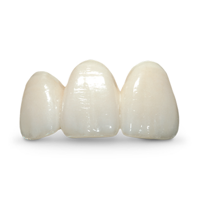
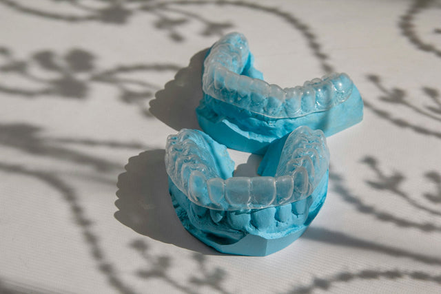
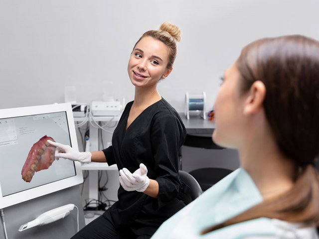
The preps were clean, the shade tab matched, and the six crowns fit the model perfectly. Then the patient smiled: the incisal plane ran uphill, the centrals looked long, and the midline sat off her face. Nothing on the Rx was wrong. The technician simply never saw the face.
Anterior work is designed on a model or a scan but judged in a smile, by a patient with expectations of her own. The records you send close that gap. Below: the records that decide an anterior crown or veneer case, why the lab needs each one, how to capture it, and when to approve.
Key Takeaways
- The AACD accreditation photo series uses 12 views per stage: 4 without retractors, 6 retracted and 2 occlusal mirror views.
- In photos of 160 people, 64% had facial asymmetry; the interpupillary line was the main horizontal reference in 88.4% of cases.
- In 158 adults, 31.64% had a dental midline more than 2 mm off the facial midline.
- On video, continuous gingival display appeared in 81% of women and 62% of men, against 35% and 21% on still photos.
- Ivoclar's current IPS e.max Press minimums: 0.3 mm for thin veneers, 0.6 mm for veneers, 1.2 mm circumferential and 1.5 mm incisal for anterior crowns.
- Clinicians rated 43.8% of anterior restorations satisfactory; the patients themselves rated 67.6% satisfactory.
Quick Answer
Send a standard photo series, a full-face photo that shows the interpupillary line and facial midline, a short smile video, the target and stump shades, a scan or model of the approved provisionals, a bilateral bite plus a protrusive record, preps measured against your material's minimum thickness, written characterization notes and the patient's requests in her own words. Approve shape in the provisionals and color at try-in, before bonding.
What Records Does the Lab Need for an Anterior Case?
| Record | Why the lab needs it | How to capture it |
|---|---|---|
| Photo series | Proportions, gingival levels, incisal plane and embrasures a model cannot show | Full face, smile, retracted views with teeth slightly apart, 1:1 close-ups, occlusals |
| Facial reference | Sets the midline and incisal plane to the face, not the model | Full-face smile, camera level, head upright |
| Smile video | Shows the spontaneous smile and speech, which posed photos understate | A few seconds of talking and laughing |
| Shade and stump shade | Choice of ingot, opacity and layering | Tabs photographed before the prep; stump shade on the prep |
| Pre-op scan | The starting shape and what you changed | Full-arch scans before preparation |
| Approved provisionals | The length, contour and phonetics the patient accepted | Scan or impression of the provisionals in place, plus photos |
| Bite and protrusive record | Incisal edges and lingual contours that clear in function | Bilateral bite in maximum intercuspation and a protrusive record |
| Prep data | Room for the material you prescribed | Reduction measured through a guide; clear margins |
| Characterization notes | Halo, translucency, texture, mamelons, staining | Written on the Rx, ideally drawn on a printed photo |
| Patient expectations | What "natural" or "whiter" means to this patient | Requests in the patient's words |
Which Photos Should You Send?
The American Academy of Cosmetic Dentistry (AACD) requires 24 views for an accreditation case, 12 before and 12 after treatment, at magnifications of 1:10, 1:2 and 1:1: a full face; a natural smile from the front and both sides; retracted views with the teeth slightly apart from the same three angles; maxillary anterior close-ups from the same three angles; and maxillary and mandibular occlusal views with a mirror. Parting the teeth, AACD notes, allows "evaluation of incisal plane and incisal embrasures."
Views in the AACD's standard series for each stage of a cosmetic case (AACD, A Guide to Accreditation Photography).
A single crown rarely needs all 12; veneer cases do. Posed photos also understate the smile: in 122 men filmed smiling, posed smiles showed up to 30% less tooth in the premolar and molar area than spontaneous ones, and the authors propose video (Van der Geld et al., 2008).
How Do You Give the Lab a Facial Reference?
Interpupillary line and facial midline
In a photographic study of 160 people, 64% had some facial asymmetry; the interpupillary line was the main horizontal reference in 88.4% of cases, the intercommissural line second, and the eye detected loss of parallelism down to about 1 degree (Margossian et al., 2021).
Share of 158 adults with at least one deviation in midline position, midline inclination or occlusal plane, measured against the bipupillary line and facial midline (Jiménez-Castellanos et al., 2016).
In that study, 31.64% had a midline more than 2 mm off the facial midline and 25.9% an occlusal plane inclined more than 2 degrees. Copy a canted dentition tooth to tooth and the cant carries into the new restorations.
How much shows? Orthodontists and general dentists rated a 1 mm incisal plane cant less esthetic, lay people only at 3 mm, and all groups noticed a 2 mm discrepancy in incisor angulation (Kokich et al., 1999).
Smile line
In Tjan's survey of 454 dental and dental hygiene students, 11% had a high smile, showing the full incisor length and a band of gingiva; 69% an average smile, showing 75% to 100% of the incisors; and 20% a low smile (as summarized by Zachrisson, 1998). Grading each tooth high, average or low was reliable against measurement (Van der Geld et al., 2011). The smile line tells the lab how much the cervical third and gingival zeniths will show; orthodontists and lay people rated 3 mm of gingival display unattractive (Kokich et al., 2006).

How Should You Send Shade and Characterization?
Take the shade before the prep, photograph two or three tabs in the plane of the teeth and record the stump shade; the method and evidence are in our guide to shade photos and stump shade.
AACD's accreditation criteria list incisal translucency, surface color characterizations, surface finish and luster under micro esthetics. Write each effect and how strongly: incisal halo, incisal translucency, mamelons, white spots, cervical chroma, texture and gloss. Say whether to match the neighbors or the tab.

Why Send the Approved Provisionals?
The provisionals are the only version of the final shape tested in the mouth: length, lip support, phonetics, midline and the patient's verdict. A scan or impression of them in place, with a bite, gives the technician that shape instead of a description of it.
Scan them after any chairside adjustment and before you remove them, in occlusion and in protrusive. If you cut a reduction guide, make it from that approved shape. On the Rx, write "copy provisionals" and list exceptions in millimeters, such as "lengthen #8 and #9 by 0.5 mm" (more in How to Fill Out a Dental Lab Rx).

Which Bite Records Does an Anterior Case Need?
Start with a bilateral bite in maximum intercuspation (see Why Crowns Come Back High). Then add a protrusive record: incisal edges and lingual contours must clear the lower incisors in protrusion, and a static bite shows one position only. In a study on casts, virtual protrusive records from an intraoral scanner detected every reference contact (100% sensitivity); lateral records, only 28% (Morsy and Hammad, 2024).
Guidance also limits material choice. Ivoclar lists absence of canine guidance as a limitation of use for minimally invasive IPS e.max crowns, and untreated bruxism for IPS e.max Press in general. Note deep bites, edge-to-edge contacts and parafunction on the Rx.
How Much Space Does Each Material Need?
| Restoration | Minimum thickness | Cementation |
|---|---|---|
| IPS e.max Press thin veneer | 0.3 mm, 0.4 mm incisal | Adhesive only; no cut-back |
| IPS e.max Press veneer | 0.6 mm, 0.7 mm incisal | Adhesive only |
| IPS e.max Press minimally invasive anterior crown | 1.0 mm | Adhesive only |
| IPS e.max Press anterior crown, stained | 1.2 mm circumferential, 1.5 mm incisal | Adhesive, self-adhesive or conventional |
| IPS e.max Press anterior crown, cut-back | Press 1.2 mm circumferential, 0.4 mm incisal, and at least half the total thickness | Adhesive, self-adhesive or conventional |
| IPS e.max ZirCAD Prime or MT anterior crown, full contour | 0.8 mm | See the IFU |
| IPS e.max Zirconia anterior crown; veneer | 0.6 mm; 0.5 mm | Crown: any of the three; veneer: adhesive only |
These are minimum thicknesses of the finished restoration, so the reduction must leave that room under the final contour, including the approved length. Other zirconia brands set their own minimums. Margins and taper: our Crown Prep Guide; veneer materials compared: Veneers: E.max vs. Zirconia vs. Feldspathic.
How Do You Set Patient Expectations?
Patients and dentists judge anterior work differently. In a Jordanian study, clinicians rated 43.8% of maxillary anterior restorations satisfactory, the patients 67.6%, and their rankings disagreed more often than they agreed (Albashaireh et al., 2009). In 90 patients with anterior fixed prostheses, patient and clinician answers agreed 47.8% to 72.2% of the time, least on whether the restoration looked natural (Alshiddi et al., 2015).
Ask what bothers the patient, write it down in her words, and agree on what will not change, such as gingival levels without periodontal treatment. Let her judge the provisionals in daylight over a few days, and photograph the version she approves.
For using a smile preview at the consultation, see AI Smile Simulation for Case Acceptance: What It Shows, What It Can't, and How to Use It.
When Should You Approve, and What Happens at Try-In?
Shape: in the provisionals
Settle length, midline and contour before the final scan or impression.
Color: at try-in, with care
Ivoclar says its Variolink Esthetic try-in pastes match the shade and translucency of the cured cement at the same layer thickness, and asks for the try-in before isolating or drying, because drying temporarily lightens the tooth. Independent lab studies are more cautious: those try-in pastes and cured cements differed significantly in color, most at 0.5 mm (Mourouzis et al., 2018), and another study recommends checking with the uncured cement in place before curing (ALGhazali et al., 2010).
Occlusion: carefully, or after bonding
For brittle ceramic restorations, Ivoclar's try-in paste instructions say to check occlusion carefully or only after final cementation. Bonding steps: How to Cement Zirconia and Bond E.max.
Anterior Crowns and Veneers at Elegant Dental Laboratory
- IPS e.max, pressed only with IPS e.max Press: full-contour and layered crowns, veneers from 0.3 mm, HO ingots for dark preparations.
- Zirconia: solid, 9-layer, solid with buccal porcelain, and copings with layered porcelain, in the brand or grade written on the Rx; also provisionals.
- Every case made in house in Brooklyn by more than 35 technicians; digital scans reviewed before production; turnaround from 5 business days on most restorative cases, rush decided case by case; up to a 7-year warranty on final restorations (policies and warranties).
- Submit a digital case or send a case: free pickup by our own drivers in all five New York City boroughs and 13 northern New Jersey counties, UPS labels anywhere. Call 877-335-5221.
FAQ
How many photos does the lab need for a veneer case?
The AACD's 12 views are a good standard, plus a short smile video.
Which facial line should the incisal plane follow?
Usually the interpupillary line, the main horizontal reference in 88.4% of cases in one study. If the eyes are not level, say which reference to use.
Why send a scan of the provisionals?
It is the shape the patient already approved. Without it, the technician designs from a description.
Do I need a protrusive record for anterior crowns?
Yes. Incisal edges and lingual contours must clear in protrusion, which a bite in maximum intercuspation does not show.
Can I trust the try-in paste for the final shade?
As a guide. Lab studies found differences between try-in pastes and cured cements, largest under thin ceramic.
When should the patient approve?
Shape in the provisionals, color at try-in, both before bonding.
Sources (19)
- American Academy of Cosmetic Dentistry. A Guide to Accreditation Photography: Photographic Documentation and Evaluation in Cosmetic Dentistry, and A Guide to Accreditation Criteria (preview), accessed 2026-10-01. PDF
- Van der Geld P, et al. Tooth display and lip position during spontaneous and posed smiling in adults. Acta Odontol Scand. 2008;66(4):207-213. doi:10.1080/00016350802060617
- Mahn E, et al. Comparing the use of static versus dynamic images to evaluate a smile. J Prosthet Dent. 2020;123(5):739-746. doi:10.1016/j.prosdent.2019.02.023
- Margossian P, et al. Determination of Facial References for Esthetic Restorative Treatment. Int J Periodontics Restorative Dent. 2021;41(1):113-119. doi:10.11607/prd.4642
- Jiménez-Castellanos E, et al. Prevalence of alterations in the characteristics of smile symmetry in an adult population from southern Europe. J Prosthet Dent. 2016;115(6):736-740. doi:10.1016/j.prosdent.2015.08.031
- Kokich VO Jr, Kiyak HA, Shapiro PA. Comparing the perception of dentists and lay people to altered dental esthetics. J Esthet Dent. 1999;11(6):311-324. doi:10.1111/j.1708-8240.1999.tb00414.x
- Tjan AH, Miller GD, The JG. Some esthetic factors in a smile. J Prosthet Dent. 1984;51(1):24-28. doi:10.1016/s0022-3913(84)80097-9
- Zachrisson BU. Esthetic factors involved in anterior tooth display and the smile: vertical dimension. J Clin Orthod. July 1998 (summary of Tjan et al.). jco-online.com
- Van der Geld P, et al. Smile line assessment comparing quantitative measurement and visual estimation. Am J Orthod Dentofacial Orthop. 2011;139(2):174-180. doi:10.1016/j.ajodo.2009.09.021
- Kokich VO, Kokich VG, Kiyak HA. Perceptions of dental professionals and laypersons to altered dental esthetics: asymmetric and symmetric situations. Am J Orthod Dentofacial Orthop. 2006;130(2):141-151. doi:10.1016/j.ajodo.2006.04.017
- Morsy N, Hammad I. Recording maximal intercuspation and border positions of the mandible with intraoral scanner using the acquisition software's multi-occlusion function. J Adv Prosthodont. 2024;16(4):221-230. doi:10.4047/jap.2024.16.4.221
- Ivoclar Vivadent. IPS e.max Press Instructions for Use, Rev. 2, 2026-01-08 (indications, limitations of use, minimum layer thicknesses, cementation). Ivoclar eIFU
- Ivoclar Vivadent. IPS e.max ZirCAD labside Instructions for Use, Rev. 3, 2026-02-11 (minimum thicknesses). Ivoclar eIFU
- Ivoclar Vivadent. IPS e.max Zirconia Instructions for Use, Rev. 0, 2026-04-27 (minimum wall thicknesses, cementation). Ivoclar eIFU
- Albashaireh ZS, Alhusein AA, Marashdeh MM. Clinical assessments and patient evaluations of the esthetic quality of maxillary anterior restorations. Int J Prosthodont. 2009;22(1):65-71. PubMed 19260431
- Alshiddi IF, BinSaleh SM, Alhawas Y. Patient's Perception on the Esthetic Outcome of Anterior Fixed Prosthetic Treatment. J Contemp Dent Pract. 2015;16(11):845-849. doi:10.5005/jp-journals-10024-1768
- Ivoclar Vivadent. Variolink Esthetic Try-In Paste Instructions for Use, Rev. 0, 2026-02-11 (shade simulation, try-in). Ivoclar eIFU
- Mourouzis P, et al. Color match of luting composites and try-in pastes: the impact on the final color of CAD/CAM lithium disilicate restorations. Int J Esthet Dent. 2018;13(1):98-109. PubMed 29379906
- ALGhazali N, et al. An investigation into the effect of try-in pastes, uncured and cured resin cements on the overall color of ceramic veneer restorations: an in vitro study. J Dent. 2010;38 Suppl 2:e78-e86. doi:10.1016/j.jdent.2010.08.013
Reviewed by Gary Fingerman, founder and CEO of Elegant Dental Laboratory, Brooklyn, New York (founded 2007). Last reviewed: October 1, 2026. Study values are from published research or manufacturer instructions; always follow the current instructions for use of the materials you prepare and bond.