The implant crown seats, the screw is torqued, and then comes the question every restoring dentist answers differently: how much should it touch? Some leave it so light that shimstock slides through in a firm clench. Others match the neighboring teeth, worried that a crown left out of the bite overloads them.
The caution has a real basis: an implant has no periodontal ligament. The evidence for any specific contact recipe is thinner than the confident rules suggest. Below: the physiology, the evidence on light contacts and overload, how to check contacts, and follow-up.
Key Takeaways
- Natural teeth move 25 to 100 µm axially in their ligament; osseointegrated implants move 3 to 5 µm.
- In 22 patients, it took 8.75 times more force to feel pressure through an implant crown than through a tooth.
- A 2021 Best Evidence Consensus Statement found a lack of evidence for any occlusal scheme that minimizes implant complications.
- In a 12-month study of 50 patients with single molar implant crowns, contacts set light at delivery did not stay light.
- Overload and bone loss remain contested: 4 of 7 clinical studies in a 2022 review found an association, all at moderate to serious risk of bias.
- In 30 subjects, the largest articulating paper mark matched the most forceful tooth only 38.3% of the time.
Quick Answer
Light, by most guidelines, but only at first. The usual target is graded contact: in light closure the natural teeth hold 12 µm shimstock while it just pulls through against a posterior implant crown; in firm closure everything holds, and posterior implant crowns get no excursive contacts. This is expert recommendation, not trial-proven. Light contacts grow heavier within months, and two studies found no outcome difference, so recheck at about 3 months and every year.
Why Do Implants Need Different Occlusion Than Teeth?
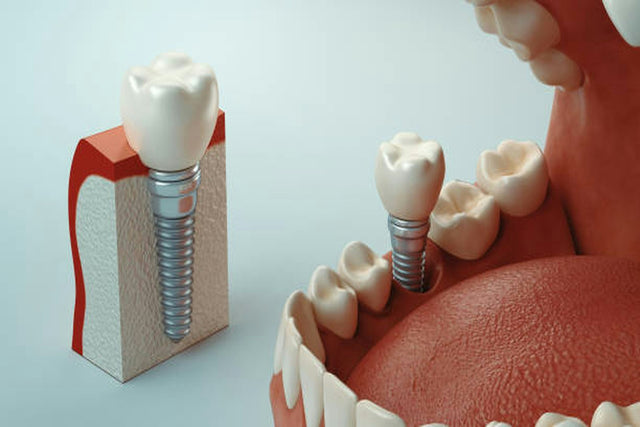
No ligament, almost no give
A tooth is suspended in a periodontal ligament that cushions load. A 2013 review puts mean axial tooth mobility at 25 to 100 µm, against 3 to 5 µm for an osseointegrated implant, with load on implants concentrating at the crestal bone (Yuan and Sukotjo, 2013). Teeth intrude slightly between light contact and firm closure; implants barely do, which is the rationale for making implant crowns slightly lighter (Stilwell, 2024).
Duller pressure sense, similar foil detection
How much higher the mean threshold for feeling pressure was on single implant crowns (100.6 g) than on natural teeth (11.5 g) in 22 patients (Hämmerle et al., 1995).
Yet when 62 patients bit on copper foil strips, the thinnest foil they detected averaged 16.7 µm between an implant crown and an opposing tooth and 14.3 µm between two natural teeth, which the authors judged equivalent (Enkling et al., 2007). Patients can often feel a high implant crown; what they lack is the ligament's cushion.

Is "Implant-Protected Occlusion" Backed by Evidence?
Implant-protected occlusion aims to protect implants by reducing occlusal force on the prosthesis. The usual advice adds shallow anatomy, a narrow occlusal table and reduced cusp inclines, yet most implant occlusion concepts rest on expert opinion, anecdotal experience and animal studies (Yuan and Sukotjo, 2013).
What reviews and consensus statements conclude
Kim and colleagues wrote in their 2005 guidelines that there is no evidence-based, implant-specific concept of occlusion, and a 2015 review found the evidence insufficient for firm guidelines (Koyano and Esaki, 2015). A 2021 Best Evidence Consensus Statement concluded that the occlusion a dentist uses for tooth- or mucosa-supported prostheses may be used on implants until compelling evidence says otherwise (Goldstein et al., 2021).
What clinical studies show about light contacts
Measured with T-Scan on 37 posterior partial fixed prostheses on implants, the implant's share of total bite force rose from 7.46% at 2 weeks to 9.87% at 3 months, while control natural teeth fell from 13.78% to 11.43%; from 6 months on, the two were statistically similar (Luo et al., 2020). In 21 patients adjusted to implant-protected occlusion, force on the implant crowns was significantly higher at 3 and 6 months than at insertion (Madani et al., 2017).

Two studies compared light and normal contacts. In 50 patients with single molar implant crowns, "light" meant a 12 µm film could be pulled out in intercuspal position. Relative force on light crowns ranged from 4.91% to 10.34% over 12 months, against 10.45% to 18.15% on normal crowns; the authors found light occlusion not stable and saw no signs of overload in either group (Zhang et al., 2024). A retrospective study of 63 implants followed for up to 5 years found the same change and no difference in marginal bone loss, proximal contact loss or technical complications (Li et al., 2026). A light contact at delivery is a starting point, not a permanent state.
Does Occlusal Overload Cause Bone Loss Around Implants?
The case that it can
In monkeys, 5 of 8 implants under excessive lateral load lost osseointegration within 4.5 to 15.5 months, while implants left to collect plaque stayed integrated but lost an average of 1.8 mm of bone (Isidor, 1996). A 2022 review of 7 clinical studies found 4 reporting a correlation between overload and crestal bone loss, all at moderate to serious risk of bias (Di Fiore et al., 2022). A 2025 review called excessive loading a critical contributor to bone loss and peri-implantitis, though it mixed clinical studies with animal work, finite element models and case reports (Mojaver et al., 2025).
The case that inflammation matters more
In dogs with healthy mucosa, 8 months of supra-occlusal contact caused no loss of osseointegration or marginal bone (Heitz-Mayfield et al., 2004). A review for the Third EAO Consensus Conference (2012) judged all 16 clinical studies it considered at high risk of bias; in animals, overload without inflammation did not harm osseointegration, while overload with inflammation increased plaque-induced bone loss (Naert et al., 2012; Klinge and Meyle, 2012). The 2017 World Workshop added that progressive crestal bone loss without soft tissue inflammation is rare (Schwarz et al., 2018).
Where the evidence agrees
Mechanical complications are less disputed: overload may lead to screw loosening or fracture and to abutment, prosthesis or porcelain fracture (Yuan and Sukotjo, 2013). Controlled contacts protect the components; healthy tissue protects the bone. See Why Implant Screws Loosen.
Which Occlusal Scheme Fits Which Situation?
| Situation | Recommended occlusal scheme | Evidence level |
|---|---|---|
| Single posterior implant crown between natural teeth | Graded contact; no excursive contacts; wide fossa, shallow cusps | Expert recommendation; cohorts show contacts intensify |
| Single anterior implant crown | Long centric freedom: shimstock just pulls through even in firm closure | Expert recommendation |
| Implant crown opposing an implant restoration | Shimstock pulls through easily in light closure, holds in firm closure | Expert recommendation |
| Posterior implant bridge | Mutually protected occlusion, anterior guidance, evenly distributed contacts, narrow occlusal table | Systematic review recommendations, evidence lacking |
| Full-arch prosthesis with cantilever | About 100 µm clearance on the cantilever; no cantilever contact in excursions | Opinion of several authors, narrative review |
| Bruxism | Same scheme plus a hard occlusal guard and annual checks | Expert recommendation; consensus suggestion without evaluable evidence |
| Immediate placement with immediate provisional | A scheme that protects the provisional during function | Consensus recommendation, 2018 |
| Fixed full-arch, edentulous maxilla | Test occlusion in an interim fixed prosthesis first | Consensus recommendation aligned with current evidence |
How Should You Check Contacts on an Implant Crown?
Shimstock for graded contact
Bausch, for example, makes a metallized shimstock film 12 µm thick. A 2024 review, illustrated with an ITI graphic, sets these targets (Stilwell, 2024):
- Light closure: natural teeth hold shimstock; it just pulls through between an implant crown and a tooth, and pulls through easily between two implant restorations.
- Firm closure: all opposing posterior teeth and implant restorations hold it.
- Excursions: no dynamic contacts on posterior implant crowns; natural teeth guide where possible.
The step from "hold" to "drag" is small: in a Harvard study of 10 participants, prototype crowns adjusted to each differed by a mean of 130 µm at the occlusal contacts (Cao et al., 2026). Adjust in small steps and recheck.
Articulating paper shows where, not how hard
Paper comes in thicknesses such as 40, 100 and 200 µm (Bausch). Under a testing machine, mark area showed no direct relationship with load (Carey et al., 2007), and in 30 subjects only about 6.7% of mark area could be explained by force (Qadeer et al., 2012).
Digital occlusal analysis
In 20 patients with implant restorations, T-Scan found high points on the implants in most patients that paper did not reveal (Chowdhary and Bukkapatnam, 2023). No indicator is neutral, though: in a lab study, the marking products generally altered the contact forces, and T-Scan was an extreme outlier (Mitchem et al., 2017). Use it alongside shimstock.
What Changes for Cantilevers and Bruxers?
Cantilevers
A 2013 review cites recommendations for minimal cantilevers alongside a systematic review that found survival comparable to prostheses without extensions, with veneer fracture and screw loosening the most common complications (Yuan and Sukotjo, 2013). On full-arch prostheses, several authors recommend about 100 µm of clearance on the cantilever (Berzaghi et al., 2025). Survival data: Cantilever Bridges.
Bruxers
Parafunction has been linked to porcelain chipping and screw loosening (Yuan and Sukotjo, 2013); see Restorations for Bruxers. A 2024 review calls a hard occlusal splint prudent with bruxism (Stilwell, 2024). The 2026 Global Consensus suggests a guard after fixed implant prostheses in the edentulous maxilla, while noting a significant lack of high-quality evidence for routine use in bruxers (Stilwell et al., 2026).
How Should You Record the Bite for the Lab?
Implant crown occlusion is set in the design from your bite record, so a wrong bite undoes everything above. Bite scans are covered in High Crowns and Open Contacts, and scan body technique in How to Scan Implants.

What Should You Do After Delivery?
- At delivery: check light closure, firm closure, then excursions; adjust the implant crown to reinstate graded contact and repolish adjusted surfaces to a high shine (Stilwell, 2024).
- At about 3 months: force on implant crowns rose significantly by 3 months and kept rising through 24 months (Luo et al., 2020), and the 21-patient study's authors call periodic adjustment necessary (Madani et al., 2017). Book this visit at delivery.
- Every year: check patient input, restoration integrity, occlusal scheme with any night guard, and spatial changes such as open proximal contacts (Stilwell, 2024). Retrievable crowns make corrections simpler; see Screw-Retained vs. Cement-Retained Implant Crowns.
Implant Restorations at Elegant Dental Laboratory
- Screw-retained and cement-retained implant restorations, custom screw-retained abutments and full-arch work, with design libraries for all major implant systems.
- Compatible scan bodies and titanium bases accepted, scan bodies available to practices, and digital scans reviewed before production (submit a digital case).
- Every case is made in house in Brooklyn by more than 35 technicians, including hard, soft and dual-laminate night guards.
- Turnaround from 5 business days on most restorative cases, rush decided case by case, and warranties of up to 7 years on final restorations and up to 20 years on screw-retained implant abutments (policies and warranties).
- Send a case: free pickup and delivery by our own drivers every business day during lab hours in all five New York City boroughs and 13 northern New Jersey counties; UPS labels anywhere. Call 877-335-5221.
FAQ
Should an implant crown be out of occlusion?
Usually not completely. Most guidelines aim for graded contact: light in light closure, full contact in firm closure.
What shimstock thickness should I use?
About 12 µm, the film thickness that defined light occlusion in the studies above.
Do light contacts on implant crowns stay light?
No. In T-Scan studies, force on implant crowns rose over the first 3 to 12 months.
Does occlusal overload cause peri-implant bone loss?
It is contested. Animal studies suggest overload mainly aggravates bone loss when inflammation is present.
Should posterior implant crowns have lateral contacts?
Recommendations say no; natural teeth should guide where possible.
Sources (30)
- Yuan JC, Sukotjo C. Occlusion for implant-supported fixed dental prostheses in partially edentulous patients: a literature review and current concepts. J Periodontal Implant Sci. 2013;43(2):51-57. doi:10.5051/jpis.2013.43.2.51
- Hämmerle CH, et al. Threshold of tactile sensitivity perceived with dental endosseous implants and natural teeth. Clin Oral Implants Res. 1995;6(2):83-90. doi:10.1034/j.1600-0501.1995.060203.x
- Enkling N, et al. Tactile sensibility of single-tooth implants and natural teeth. Clin Oral Implants Res. 2007;18(2):231-236. doi:10.1111/j.1600-0501.2006.01321.x
- Kim Y, Oh TJ, Misch CE, Wang HL. Occlusal considerations in implant therapy: clinical guidelines with biomechanical rationale. Clin Oral Implants Res. 2005;16(1):26-35. doi:10.1111/j.1600-0501.2004.01067.x
- Koyano K, Esaki D. Occlusion on oral implants: current clinical guidelines. J Oral Rehabil. 2015;42(2):153-161. doi:10.1111/joor.12239
- Goldstein G, Goodacre C, Taylor T. Occlusal schemes for implant restorations: Best Evidence Consensus Statement. J Prosthodont. 2021;30(S1):84-90. doi:10.1111/jopr.13319
- Carlsson GE. Dental occlusion: modern concepts and their application in implant prosthodontics. Odontology. 2009;97(1):8-17. doi:10.1007/s10266-008-0096-x
- Luo Q, Ding Q, Zhang L, Xie Q. Analyzing the occlusion variation of single posterior implant-supported fixed prostheses by using the T-scan system: A prospective 3-year follow-up study. J Prosthet Dent. 2020;123(1):79-84. doi:10.1016/j.prosdent.2018.12.012
- Madani AS, et al. Post-insertion posterior single-implant occlusion changes at different intervals: a T-Scan computerized occlusal analysis. J Contemp Dent Pract. 2017;18(10):927-932. doi:10.5005/jp-journals-10024-2151
- Zhang R, Hao X, Zhang K. Evaluation of two different occlusal patterns on single posterior implant-supported crowns: A 12-month prospective study of occlusal analysis. J Prosthet Dent. 2024;132(6):1277-1284. doi:10.1016/j.prosdent.2022.11.026
- Li B, et al. Comparison between light- and non-light occlusion on clinical outcomes: a retrospective study. Clin Implant Dent Relat Res. 2026;28(1):e70110. doi:10.1111/cid.70110
- Isidor F. Loss of osseointegration caused by occlusal load of oral implants. A clinical and radiographic study in monkeys. Clin Oral Implants Res. 1996;7(2):143-152. doi:10.1034/j.1600-0501.1996.070208.x
- Di Fiore A, et al. Peri-implant bone loss and overload: a systematic review focusing on occlusal analysis through digital and analogic methods. J Clin Med. 2022;11(16):4812. doi:10.3390/jcm11164812
- Mojaver S, Patel N, Sarmiento H, Fiorellini JP. Under pressure: unraveling the impact of occlusal overload on peri-implant health: a systematic review. J Prosthodont. 2025;34(8):784-795. doi:10.1111/jopr.14088
- Heitz-Mayfield LJ, et al. Does excessive occlusal load affect osseointegration? An experimental study in the dog. Clin Oral Implants Res. 2004;15(3):259-268. doi:10.1111/j.1600-0501.2004.01019.x
- Naert I, Duyck J, Vandamme K. Occlusal overload and bone/implant loss. Clin Oral Implants Res. 2012;23 Suppl 6:95-107. doi:10.1111/j.1600-0501.2012.02550.x
- Klinge B, Meyle J. Peri-implant tissue destruction. The Third EAO Consensus Conference 2012. Clin Oral Implants Res. 2012;23 Suppl 6:108-110. doi:10.1111/j.1600-0501.2012.02555.x
- Schwarz F, Derks J, Monje A, Wang HL. Peri-implantitis. J Clin Periodontol. 2018;45 Suppl 20:S246-S266. doi:10.1111/jcpe.12954
- Sheridan RA, Decker AM, Plonka AB, Wang HL. The role of occlusion in implant therapy: a comprehensive updated review. Implant Dent. 2016;25(6):829-838. doi:10.1097/ID.0000000000000488
- Stilwell C. Occlusal considerations in maintaining health of implants and their restorations. Br Dent J. 2024;236(10):773-779. doi:10.1038/s41415-024-7407-7
- Berzaghi A, Testori T, et al. Occlusion and biomechanical risk factors in implant-supported full-arch fixed dental prostheses: narrative review. J Pers Med. 2025;15(2):65. doi:10.3390/jpm15020065
- Stilwell C, Jung RE, Beuer F, Brunello G, et al. Consensus report of Group 4 of the 1st Global Consensus for Clinical Guidelines for the Rehabilitation of the Edentulous Maxilla. Clin Oral Implants Res. 2026;37(Suppl 30):S81-S107. doi:10.1111/clr.70069
- International Team for Implantology. Implant placement and loading protocols: consensus statements, ITI Consensus Conference 2018 (Gallucci G, Hamilton A, Zhou W, Buser D, Chen ST). ITI consensus database
- Bausch. Arti-Fol articulating films, including Arti-Fol metallic 12µ shimstock film (product page, accessed October 1, 2026). bauschpaper.com
- Bausch. Articulating papers 40µ, 100µ and 200µ (product page, accessed October 1, 2026). bauschpaper.com
- Cao H, Lopez D, Lee JD, Lee SJ. Quantitative analysis of occlusion in posterior single-implant supported restorations: a clinical study. J Dent. 2026;164:106233. doi:10.1016/j.jdent.2025.106233
- Carey JP, Craig M, Kerstein RB, Radke J. Determining a relationship between applied occlusal load and articulating paper mark area. Open Dent J. 2007;1:1-7. doi:10.2174/1874210600701010001
- Qadeer S, et al. Relationship between articulation paper mark size and percentage of force measured with computerized occlusal analysis. J Adv Prosthodont. 2012;4(1):7-12. doi:10.4047/jap.2012.4.1.7
- Chowdhary R, Bukkapatnam S. Comparative evaluation of implant-protected occlusion in partially edentulous fixed restoration using qualitative and quantitative assessment: a prospective clinical trial. Indian J Dent Res. 2023;34(4):396-400. doi:10.4103/ijdr.ijdr_191_23
- Mitchem JA, Katona TR, Moser EAS. Does the presence of an occlusal indicator product affect the contact forces between full dentitions? J Oral Rehabil. 2017;44(10):791-799. doi:10.1111/joor.12543
Reviewed by Gary Fingerman, founder and CEO of Elegant Dental Laboratory, Brooklyn, New York (founded 2007). Last reviewed: October 1, 2026. Most implant occlusion guidelines are expert recommendations; use them alongside your own clinical judgment.